FACILITIES

Pentacam-Gold Standard in Anterior Segment Tomography

Understanding the shape and thickness of the cornea is important to any refractive procedure. The Pentacam is a diagnostic test that analyzes and measures the cornea, the clear front surface of the eye where light rays enter.

When considering laser VISION CORRECTION or cataract surgery, this advanced diagnostic instrument is a real advantage over all other ocular diagnostic equipment available today.

Optical Biometer AL-Scan

IOL Station Software for the OPD-Scan: This wavefront a berrometry system determines corneal, internal, and total-eye aberrations and provide the optimal axis of alignment for Toric IOLs

In 10 seconds, six values for cataract surgery are measured

  • Axial length
  • Corneal curvature radius
  • Anterior chamber depth
  • Central corneal thickness
  • White-to-white distance
  • Pupil size

S L-D7 Digital-Ready Slit Lamp

The Topcon SL-D7 with its optional Digital Photo Attachment is the only digital photo slit lamp with a flash-through-the-slit illumination system. This allows for unsurpassed clarity and image excellence in slit lamp photography.

The SL-D7 can be interfaced with the optional DC-3 integrated digital camera for 8 megapixel imaging or the Nikon photo attachment for an outstanding 12 megapixels of resolution.

3D Maestro OCT from TOPCON

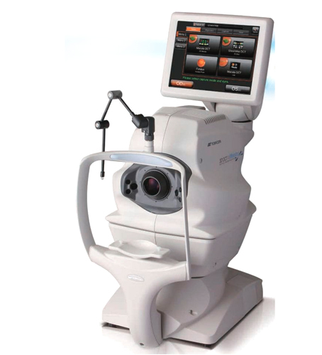
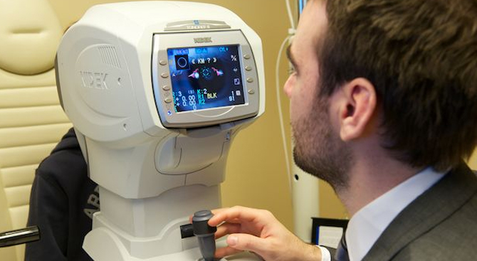
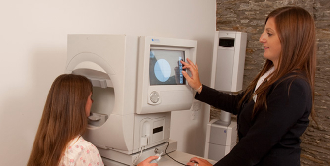
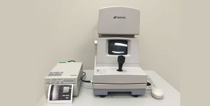
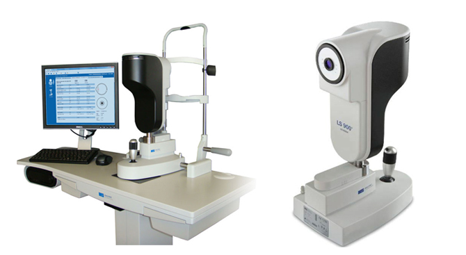
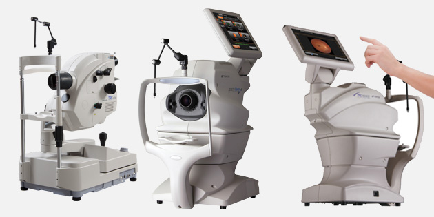
The Topcon 3D OCT-2000 System is the first Spectral Domain OCT system to incorporate a high resolution fundus camera and a user friendly color touch screen display The easy-to-use, intuitive FastMap™ software enables dynamic viewing of the OCT data, providing 3D, 2D and fundus images simultaneously. Quick, safe and informative–this new Eye scanner is a glimpse into the future of patient care.


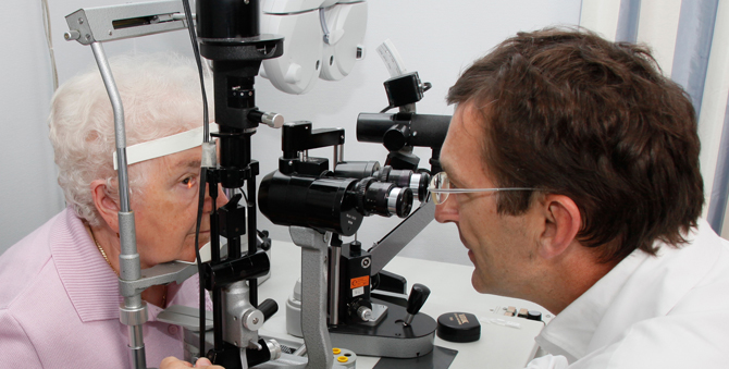
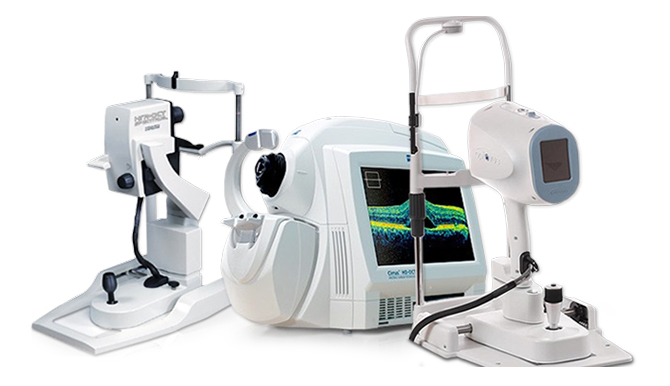
It gives live cross sections magnified slices of various structures of the eye, giving a better insight in the treatment of the eye disease.We can look for the early stages of diseases like glaucoma, diabetic retinopathy and macula edema, all of which have the potential to change your sight.

This is a scanning machine which helps in doing two tests

  • A scan biometry – This is a test to calculate the length of the eye ball. This is a very important measurement for accurate calculation of IOL power.
  • Sonography of the eye or B scan – This is test to know the status of the retina inside the eye where direct visualisation of the retina and optic nerve is not possible eg in patients with dense cataracts, corneal opacity or in children and patients with eye injury.

Applanation Tonometer

This is a gold standard for measuring for measuring pressure inside the eye or Intraocular pressure.It measures pressure by force required to flatten a constant area of the cornea.

Auto Refractometer

  • This instrument is used for computerised testing of refractive power of the eye.

Keratometer

This is used to measure the curvature of front surface of the cornea. This reading is helpful in measuring the IOL power before cataract surgery.It is also used for perfect fitting of contact lenses.

Non Contact Tonometer

  • It optically detects the momentary state of the cornea (applanated by air pressure) and measures intraocular pressure without touching the cornea.

Perimeter Or Humphrey Field Analysed 750

  • The Gold Standard in glaucoma diagnosis and management offers the utmost in clinical confidence
  • Advanced analysis tools backed by decades of research and clinical validation
  • SITAT SWAP software reduces blue-yellow threshold test time to just 3 to 6 minutes, providing a clinically practical tool for early glaucoma detection

Slit Lamp

  • Our institute is equipped with 2 different slit lamps by Takagi and Appasamy. This is a type of microscope with attached light which allows doctor to examine eye under high magnification. It is primarily used to view the anterior structures of the eye like cornea, lens and iris. However with the use of specialised lenses, it is also possible to see the vitreous, retina and optic nerve with this.

Specular Microscope. Sp 2000p

  • It is used for photomicroscopic visualisation and qualitative and quantitative documentation of corneal endothelial cells. These cells are required for maintaining the transparency of cornea. This test is done preoperatively in cases of cataract to know the status of cornea and predict visual outcome after surgery.

Orbscan Ii (USA) Or Corneal Topographer

  • The Orbscan II® is the most highly sophisticated corneal topographer on the market. Every LASIK evaluation must include corneal topography, which is a high resolution analysis of the cornea. Most corneal topography units in use today analyze the front surface of the cornea only. The Orbscan II® used at The Doctor eye Institute analyzes both the front and back surfaces of the cornea as well as determining the corneal thickness. This additional information can be crucial in determining whether a patient can have LASIK surgery performed on them safely.


    Experienced LASIK surgeons would refuse to perform LASIK without an Orbscan II® as part of the pre-operative evaluation.

Zeiss (Germany) Nd Yag Laser

  • This laser is used for 2 purposes :

    • It is used for doing capsulotomy for the treatment of secondary cataract or posterior capsular opacification which develops in about 50 % of the patients after cataract surgery.
    • It is also used for doing peripheral iridectomy in patients with angle closure glaucoma. It is a condition of increased pressure inside the eye caused by sudden blockage of circulation of fluid within the eye.

Axis Ii A Scan With Ultrasonic Pachymeter

  • This instrument is used for calculating axial length of the eye which helps in determining IOL power before cataract surgery. The addition of ultrasonic pachymeter with this machine helps in accurate measurement of corneal thickness for proper corneal evaluation and patient selection before keratorefractive procedures such as LASIK.

Zeiss Green Laser Or Double Frequency Yag Laser

  • LASER It is used for treatment of disease like Diabetics Retinopathy, Vascular Occlusions and peripheral retinal degenerations.


    It is fully equipped with three delivery systems:

    • Slit Lamp delivery system
    • Laser indirect Ophthalmoscope
    • Endolaser delivery System

Lenstar

  • Intraocular Lenses (IOLS) are the lenses that are placed inside the eye after removing the cataractous lens.

    Measurement of the entire eye in one scan

    The Lenstar is the first optical biometer on the market providing the surgeon with all measurements necessary to take full advantage of the latest IOL prediction methods like Holladay II or Olsen.

    The measurement includes corneal thickness, anterior chamber depth, lens thickness, axial length, keratometry, white-to-white distance, pupillometry, eccentricity of the visual axis and retinal thickness* at the point of fixation (macula).

Topcon Imagenet Retinal Imaging System

  • It is used for Fundus Fluoroscien Angiography (FFA) and Fundus Photography. This is connected to image net software for easy Archiving and retrieval of Photographs and instant reporting

Optical Coherence Tomography

  • Quick, safe, simple and informative–this new retina scanner is a glimpse into the future of patient care It is a non-invasive technology used for imaging the retina, the multi-layered sensory tissue lining the back of the eye. OCT, the first instrument to allow doctors to see cross-sectional images of the retina.


    It is also used for imaging, Diagnosing and monitoring the optic nerve and retinal nerve fiber layer, the areas of the eye damaged by glaucoma. OCT is very useful for the measurement of corneal thickness (pachymetry) as well as for mapping of the cornea which has important diagnostic and surgical applications